Our cells are very important to our longevity. They are also very, very tiny. That makes it pretty hard to study them in detail. If we want to understand how they work – and maybe take some steps to stop them from deteriorating with age – first we need to be able to see them. With modern technology, that’s becoming easier and easier.
This story begins back in the 17th century, when scientists discovered cells for the first time. It wasn’t until the 20th century, however, when electron microscopy became possible and they were really able to dig into the organelles and proteins and other parts that make a cell. An organelle is to a cell what an organ is to the larger body. That’s where it gets its name.
As the 20th century progressed, there were even more new techniques and technologies. X-ray crystallography was a big one, providing a closer look at the myoglobin protein. Then came the 21st century. In just the last few years, we’ve seen the advent of nuclear magnetic resonance and cryo-electron microscopy. Each new method meant scientists could visualize even more of the cell’s complexity.
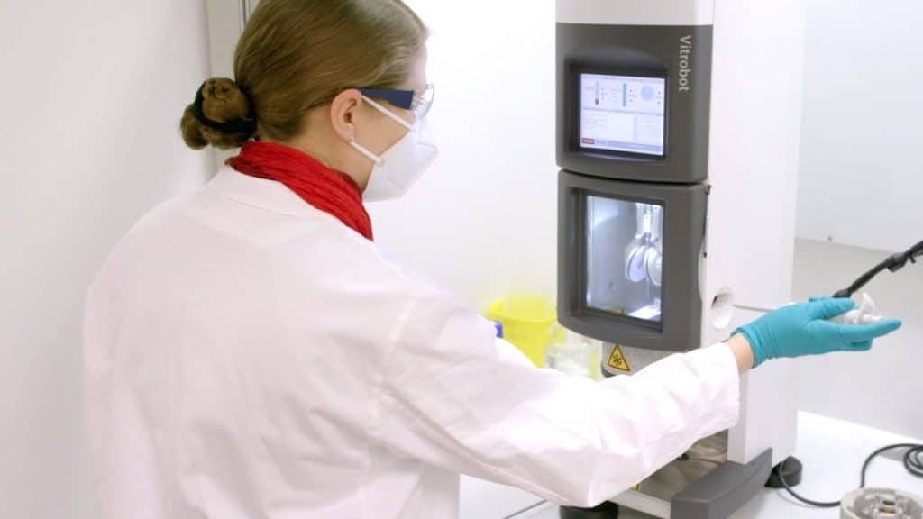
Now there’s cryo-electron tomography, or cryo-ET (CET). First, vitreous ice is used to freeze a very thin slice of cell. Then, pictures can be taken with an electron microscope. These pictures will be tilted in different ways and taken at different angles. A computer then reconstructs these images in high-resolution 3D to show every cell component in the maximum degree of complexity. Artificial intelligence even allows scientists to predict structures for the proteins they haven’t discovered yet.
CET images have a resolution of 1 to 2 nanometers, which is incredibly strong. They have other advantages, too. Other forms of imaging require the cells to be dehydrated or undergo chemical fixation first, which can be damaging processes. CET needs neither, although its functioning is complicated by the fact that it needs a high-vacuum environment to work. It’s also a fairly laborious, time-consuming job.
What we need to do next is figure out which parts of the cell we want to focus on and continue to make use of the other new and ever-improving technologies, like the fluorescent tags of CLEM, that will hopefully make things even easier. By studying the smallest parts of organisms, perhaps we’ll better understand the organism as a whole.