If you want to know if there’s something wrong in the brain, it seems obvious that you need to take a look at the brain itself. There are some particularly annoying conditions that don’t show up on any kind of scan, but when the issue is structural, that’s when neuroimaging comes into play.
You’ve probably heard the names, or at least the acronyms, of the most common forms of neuroimaging, such as MRIs, CAT scans and PET scans. You wouldn’t think that just taking a picture of one organ could be so complicated, but that’s the brain for you. It works in many different ways, and we still barely understand even the most basic of them.
Angelo Mossi was the first person to attempt modern neuroimaging back in the 1880s. He tried to measure how blood flow changed in relation to brain activity. Then, in the early 1900s, pneumoencephalography became a thing. That’s when you drain the cerebrospinal fluid surrounding the brain and replace it with air. Yes, it’s as painful and unpleasant as it sounds. At the time, though, it made the brain clearer on x-rays.
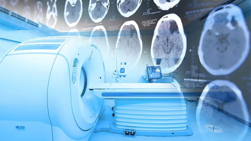
It wasn’t until the 1970s that neuroimaging as we know it today began. First, there were computer tomography (CT), or computer axial tomography (CAT) scans. You lie in a tube that takes x-rays of different parts of the body at different angles. This allows the creation of cross-sectional images showing slices of those body parts, which can include the brain. This might show evidence of strokes, tumors, bleeding and other trauma.
Then there’s the MRI, which stands for magnetic resonance imaging. As the name suggests, it uses magnetic fields to scan the body. This means you can’t have any metal on when you go into the machine, which is awkward for people with metal pins, plates or pacemakers. It gives a better view of the soft tissue than a CT scan and may reveal anything from cancer to dementia to epilepsy. Functional magnetic resonance imaging shows blood flow.
Positron emission tomography (PET) scans use chemical compounds called radiotracers. By tracking their radioactive signals, we can trace blood flow to the brain, as well as activity in the dopamine, serotonin and opioid receptors.
These are only three of many forms of neuroimaging. Scientists are constantly improving them and developing new methods on the never-ending quest to understand the brain.